
Sea urchin skeleton (stereom)
Cidaris rugosa
zenodo-10549062-732nm-inner-interambulacral-plate-lattice-x384y640z256-n128
Grade and method
- Grade
- Grade A: volumetric source.
- Method
- V: volumetric re-mesh (marching cubes on a licensed volume)
- Mesh method
- B2: marching cubes (Lewiner, level 0.5) on the cleaned binary volume; every vertex is then moved along its marching-cubes edge to the linear crossing of the grey-level threshold (grey field forced to the sign of the cleaned mask, margin 0.01 x the p0.1 to p99.9 grey span); the triangles are those of the binary mesh
- Structure
- echinoid skeletal stereom (inner interambulacral plate)
- Taxon
- Cidaris rugosa
- Imaging
- micro-CT
- Zone
- interior
Renders
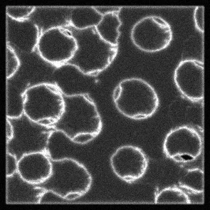
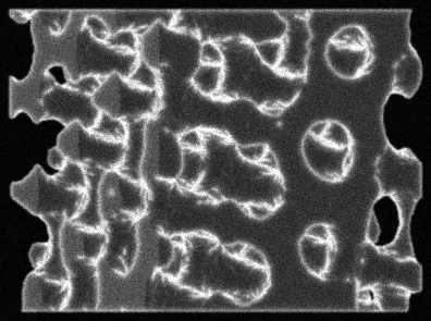
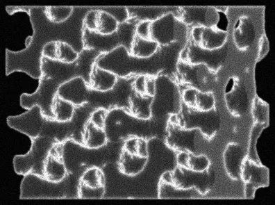
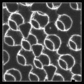
Licence and source
- Licence
- CC0 1.0 Universal
- Attribution
- Jessop, Anna-Lee (2024). Micro-CT data from the skeleton of the sea urchin Cidaris rugosa at four different resolutions. https://doi.org/10.5061/dryad.wdbrv15vf. Licence: CC0-1.0.
- Source DOI
- https://doi.org/10.5061/dryad.wdbrv15vf
- Source record
- zenodo-10549062
- File
- 732nm_inner_interambulacral_plate.tif
- Panel
- 732nm_inner_interambulacral_plate.tif#x384y640z256n128
- SHA-256 of the original
- 59d2b14b489ad3ae13c31aaabcb46d1161a8fc1c04c726123d6dd8e572438401
- Retrieved
- 2026-11-14 06:08:41 UTC
Parameters and their sources
Every parameter is tagged measured, literature or estimated.
| Parameter | Value | Unit | Uncertainty | Tag | Source detail |
|---|---|---|---|---|---|
| Voxel size | 0.732 | µm | literature | Record README (Micro-CT data from the skeleton of the sea urchin Cidaris rugosa); voxel size statement | |
| Cube side | 93.696 | µm | measured | side voxels x voxel size; method version entries-1; panel 732nm_inner_interambulacral_plate.tif#x384y640z256n128 | |
| Solid fraction, final | 0.341608 | measured | segmentation: global otsu threshold on a deterministic sample; grey > t is solid; method version entries-1; panel 732nm_inner_interambulacral_plate.tif#x384y640z256n128 | ||
| Solid fraction, raw | 0.342475 | measured | segmentation: global otsu threshold on a deterministic sample; grey > t is solid; method version entries-1; panel 732nm_inner_interambulacral_plate.tif#x384y640z256n128 | ||
| Threshold | 30,868.7 | grey level | measured | segmentation: global otsu threshold on a deterministic sample; grey > t is solid; method version entries-1; panel 732nm_inner_interambulacral_plate.tif#x384y640z256n128 | |
| Segmentation method | global otsu threshold on a deterministic sample; grey > t is solid | measured | segmentation: global otsu threshold on a deterministic sample; grey > t is solid; method version entries-1; panel 732nm_inner_interambulacral_plate.tif#x384y640z256n128 | ||
| Limit: cut-face share | 0.35 | fraction of mesh area | estimated | limit adopted on 2026-11-19 for the cut-face share and solid fraction of an entry | |
| Limit: final solid fraction | 0.6 | fraction of voxels | estimated | limit adopted on 2026-11-19 for the cut-face share and solid fraction of an entry | |
| Limit: zero voxels in a greyscale cube | 0.01 | fraction of voxels | estimated | default rule: at most 1 % of a greyscale cube is exactly 0 |
Mesh
Checked on the 3MF file reloaded from disk.
- Triangles
- 500,196
- Vertices
- 249,926
- Extent (µm)
- 93.696 × 93.696 × 93.696
- Components
- 1
- Genus
- 87
- Watertight
- yes
- Boundary edges
- 0
- Non-manifold edges
- 0
- 3MF unit
- micron
- 3MF file size
- 4.8 MB
- SHA-256 of the 3MF
- 11793b90ce370c2ab85109f6fc6b06bd2316eb33289d5e6fdd644d1874fa2ce9
Region
- Zone
- interior
- Rule
- non-overlapping cubes of 128 voxels on a lattice, in a seeded random order (numpy default_rng). Specimen envelope: the segmented volume reduced to cells of 8 voxels (a cell is solid when at least 0.05 of its voxels are solid), closed 4 steps, the outside being the empty cells connected to the volume border and everything else the envelope. A cube wholly inside the envelope is interior, one holding envelope and outside cells is surface, one wholly outside is skipped. Every interior cube with raw solid fraction in [0.25, 0.85] and every surface cube in [0.1, 0.85] is taken (or the first N of them in the seeded order); a greyscale cube with more than 0.01 zero voxels is skipped
- Origin (voxels)
- 384, 640, 256
- Side (voxels)
- 128
Filter values
- Rule
- keep a cube only if its final (cleaned) solid fraction is at most 0.6 and its cut-face share (mesh area lying in the six crop planes, all three vertices within 0.01 of the smallest voxel size of a plane, divided by the total mesh area) is at most 0.35
- Final solid fraction
- 0.3416 (limit 0.6)
- Cut-face share
- 0.1611 (limit 0.35)
Print scale
Suggested print scale ×500 (printed length divided by true length), giving 46.85 × 46.85 × 46.85 mm. This is a suggestion only: scale the mesh yourself. The entry is one 3MF in micrometres; there is no separate print file.
Downloads
On Hugging Face, release tag v0.2.0.
- 3MF mesh (micron) (4.8 MB; SHA-256 11793b90ce370c2ab85109f6fc6b06bd2316eb33289d5e6fdd644d1874fa2ce9)
- params.json
- Render PNG, face
- Render PNG, oblique30
- Render PNG, oblique60
- Render PNG, girdle
- Entry folder
- metadata.jsonl (set)
- ATTRIBUTION.md (set)
- CITATION.cff (set)


How to cite
Sunnyday Technologies (2026). Evolved Microstructure Atlas: cidaris-stereom, version 0.2.0. https://huggingface.co/datasets/evolved-microstructure-atlas/cidaris-stereom. Entry zenodo-10549062-732nm-inner-interambulacral-plate-lattice-x384y640z256-n128.
Cite the source as well:
Jessop, Anna-Lee (2024). Micro-CT data from the skeleton of the sea urchin Cidaris rugosa at four different resolutions. https://doi.org/10.5061/dryad.wdbrv15vf. Licence: CC0-1.0.